Your Eye

Your Eye

Your Eye

Your Eye

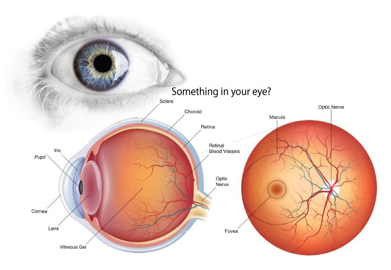
EXAMINING THE INTERIOR OF THE EYE: Your eye is a specialized sense organ capable of receiving visual images, busily absorbing light and energy that brings you vision. If you’d like to know more about what’s in your eye, this brief synopsis can help. Any additional questions, just ask Dr. Plous.
Sclera – Supporting structure of the eye made up of connective tissue. The sclera connects with the cornea, and is called the “white” of the eye.
Iris – The colored part of the eye that controls the amount of light that reaches the retina. The iris does this by controlling the size of the pupil.
Pupil – The opening within the Iris that allows light to pass into the eye. The larger it is the more light enters the eye.
Cornea – The clear front part of the eye that connects with the sclera and serves as the primary refractive structure of the eye. This is the window into the eye.
Lens – A structure composed of proteins that changes shape to focus light on the retina.
Vitreous Gel – The optically transparent gelatinous substance that fills the area between the lens and retina. The vitreous helps maintain the eye’s shape.
Retina – The light sensitive inner lining of the back of the eye. The retina converts light into electrical signals that are then transmitted to the brain through the optic nerve.
Optic Nerve – Located at the back of the eye, it transmits electrical signals created by the retina to the brain where they are experienced as images.
Macula – The central area of the retina that provides your central vision. You use your macula when you are viewing something straight ahead. The structures and cells within the macula are responsible for highly detailed, sharp, vision.
Fovea – Located in the center of the macula and the area where fine vision and color perception are the greatest.
Retina Blood Vessels – Bring oxygen and nourishment to the retina.
Choroid – The highly vascular layer between the sclera and the retina that provides oxygen and nourishment to the retina.
What are Common Retinal Diseases?
- Retinal tear. A retinal tear occurs when the clear, gel-like substance in the center of your eye (vitreous) shrinks and tugs on the thin layer of tissue lining the back of your eye (retina) with enough traction to cause a break in the tissue. It’s often accompanied by the sudden onset of symptoms such as floaters and flashing lights.
- Retinal detachment. A retinal detachment is defined by the presence of fluid under the retina. This usually occurs when fluid passes through a retinal tear, causing the retina to lift away from the underlying tissue layers.
- Diabetic retinopathy. If you have diabetes, the tiny blood vessels (capillaries) in the back of your eye can deteriorate and leak fluid into and under the retina. This causes the retina to swell, which may blur or distort your vision. Or you may develop new, abnormal capillaries that break and bleed. This also worsens your vision.
- Epiretinal membrane. Epiretinal membrane is a delicate tissue-like scar or membrane that looks like crinkled cellophane lying on top of the retina. This membrane pulls up on the retina, which distorts your vision. Objects may appear blurred or crooked.
- Macular hole. A macular hole is a small defect in the center of the retina at the back of your eye (macula). The hole may develop from abnormal traction between the retina and the vitreous, or it may follow an injury to the eye.
- Macular degeneration. In macular degeneration, the center of your retina begins to deteriorate. This causes symptoms such as blurred central vision or a blind spot in the center of the visual field. There are two types — wet macular degeneration and dry macular degeneration. Many people will first have the dry form, which can progress to the wet form in one or both eyes.

All Eye
Care Services

Keep
In Touch
contact information
office hours
- Monday:8:00 AM - 5:00 PM
- Tuesday:8:00 AM - 5:00 PM
- Wednesday:8:00 AM - 5:00 PM
- Thursday:8:00 AM - 5:00 PM
- Friday:8:00 AM - 5:00 PM
- Saturday:Closed
- Sunday:Closed
© 2023 Gulf Coast Retina Center. All Rights Reserved.
Powered by: